
By Mai P. Hoang, Martin C. Mihm Jr.
Melanocytic Lesions: A Case dependent procedure presents a concise but accomplished guide on how one can diagnose universal in addition to troublesome and not easy melanocytic lesions. within the first eleven chapters, each one entity is illustrated by means of a precise case; by way of dialogue of the way the analysis is reached, of the histologic differential diagnoses and of diagnostic pitfalls and ends with bulleted instructing issues. Pertinent and recent references are integrated on the finish of every bankruptcy. The latter chapters hide present microstaging and category of cancer, ancillary concepts together with immunohistochemistry in addition to to be had molecular assays and molecular exact remedy. All figures and glass slides of the mentioned situations are hosted on-line for simple viewing and entry. Melanocytic Lesions: A Case established strategy will serves as an invaluable source for pathologists, dermatologists and researchers facing melanocytic lesions.
Read Online or Download Melanocytic Lesions: A Case Based Approach PDF
Similar dermatology books

The main complete resource at the topic, this moment version is totally revised and accelerated to bare the newest advances, applied sciences, and tendencies in hair and hair care science-tracking the advance of hair care items, the emergence of recent regulatory practices, and the most recent tools in product security and efficacy overview.

Erythema - A Medical Dictionary, Bibliography, and Annotated Research Guide to Internet References
It is a 3-in-1 reference ebook. It offers a whole scientific dictionary masking hundreds and hundreds of phrases and expressions in relation to erythema multiforme. It additionally supplies huge lists of bibliographic citations. ultimately, it presents info to clients on find out how to replace their wisdom utilizing a number of net assets.
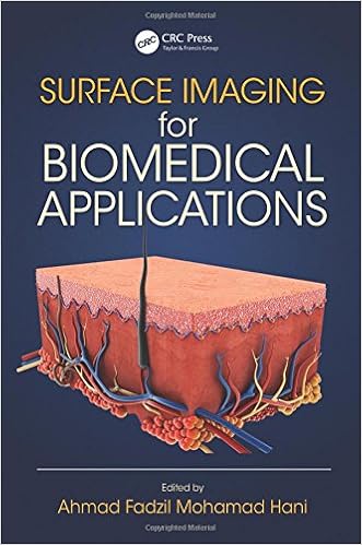
Surface Imaging for Biomedical Applications
In response to clinic medical trials studying using sign and picture processing suggestions, floor Imaging for Biomedical functions bridges the space among engineers and clinicians. this article bargains an intensive research of biomedical floor imaging to scientific practitioners because it pertains to the prognosis, detection, and tracking of dermis stipulations and ailment.

Adverse Cutaneous Drug Reactions to Cardiovascular Drugs
Antagonistic cutaneous drug reactions (ACDR) are one of the such a lot widespread occasions in sufferers receiving drug treatment. Cardiovascular (CV) medicines are an immense crew of gear with strength chance of constructing ACDR specifically in aged as advertising of extra new medicinal drugs and their prescription proceed to extend.
- Fitzpatrick's Dermatology in General Medicine
- Goodheart's Same-Site Differential Diagnosis: A Rapid Method of Diagnosing & Treating Common Skin Disorders
- Principles and practice of burn surgery
- Ethnic Dermatology: Clinical Problems and Skin Pigmentation
Extra info for Melanocytic Lesions: A Case Based Approach
Sample text
A variety of epidermal changes can be seen overlying a dermal nevus, including papillomatous hyperplasia resembling seborrheic keratosis and effacement or elongation of the rete ridges. Papillomatous hyperplasia in nevi has been attributed to estrogen effect (Morgan et al. 1995). Diagnosis Dermal nevus Comment The hallmark of the dermal nevus is a picture of a lesion that at low magnification has pale nuclei and very tiny nucleoli. These cells in the acquired lesion tend not to infiltrate in any significant way into the reticular dermis.
Clinically it resembles the other common acquired nevi (Schrader and Helwig 1967; Lewis 1969; Goette and Doty 1978). The balloon cell is a variant of the nevus cell that has a unique cytoplasm that appears clear after processing and fixation with resulting dissolution of the materials. Balloon cell alteration is found in approximately 2 % of nevi 39 and is attributed to degenerative changes (Goette and Doty 1978). This cytoplasmic change must be seen in over 50 % of the cells to qualify the lesion to be a balloon cell nevus (Schrader and Helwig 1967).
11). Toward the deeper portion of the lesion in the deep penetrating aspect, the cells all have thin delicate nuclei and are associated with fine grayish reticulum fibers that intermingle with the eosinophilic reticular dermal collagen bundles (Fig. 12). Mast cells are easily observed scattered amidst the involved area. There is definitely a plexiform pattern visible especially on low power as the cells follow the neurovascular bundles (Fig. 13). thin nuclei and scant cytoplasm. Evidence of their type-C character is the neurotization phenomenon that is present throughout the entire lesion – that is, the presence of their grayish cytoplasm as well as the presence of mast cells.